Pelvis Muscles Mri Anatomy
Pelvis Muscles Mri Anatomy. Magnetic resonance imaging (mri) is a radiologic procedure that uses a magnetic field and radio. The muscle that forms the major part. The lateral superficial muscles, the transversus and external and internal oblique muscles, originate on the rib cage and on the pelvis (iliac crest and inguinal ligament) and are attached to the anterior and posterior layers of the sheath of the rectus.
Multisystem selenoprotein deficiency trunk common: Learn about anatomy muscles pelvis with free interactive flashcards. Normal anatomy, variants and checklist. The lateral superficial muscles, the transversus and external and internal oblique muscles, originate on the rib cage and on the pelvis (iliac crest and inguinal ligament) and are attached to the anterior and posterior layers of the sheath of the rectus. Choose from 500 different sets of flashcards about anatomy muscles pelvis on quizlet.

We have 10 images about mri pelvis muscle anatomy including images, pictures, photos, wallpapers, and more.
In the back the posterior superior iliac spines are surrounded by muscles and flank fat. A pelvis mri (magnetic resonance imaging) scan is an imaging test that uses a machine with powerful magnets and radio waves to create pictures this mri pelvis cross sectional anatomy tool is absolutely free to use. Anatomy of the human body for artists course. Pelvic floor muscles that are located wholly within the pelvis. Key facts about the muscles of the pelvic floor. Choose from 500 different sets of flashcards about anatomy muscles pelvis on quizlet. We have 10 images about mri pelvis muscle anatomy including images, pictures, photos, wallpapers, and more. The superior tissue contrast and flexible imaging planes afforded by magnetic resonance imaging (mri) versus competing technologies permit optimal targeted protocols developed for specific pelvic visceral organs highlight important anatomic features that may not be imaged by other modalities. Included within the chart are gorgeous illustrations of the pelvic diaphragm, sphincter muscles, gluteus maximus. This mri pelvis cross sectional anatomy tool is absolutely free to use. Multisystem selenoprotein deficiency trunk common:
In the back the posterior superior iliac spines are surrounded by muscles and flank fat. Mri anatomy and positioning series module 5: This section of the website will explain large and minute details of axial male pelvis use the mouse scroll wheel to move the images up and down alternatively use the tiny arrows (>>) on both side of the image to move the images. Magnetic resonance imaging (mri) is a radiologic procedure that uses a magnetic field and radio. Anatomy and pathology of the male pelvis by magnetic resonance imaging. Mri pelvis anatomy | free male pelvis axial anatomy from mrimaster.com start studying mri anatomy pelvis. Related online courses on physioplus. ► shoulder ► elbow ► wrist ► finger ► thumb. The ligament around this joint relaxes during pregnancy. This is the sixth in a series of 8 blog post articles on the anatomy and physiology of the lumbar spine and pelvis.

Not only mri pelvis muscle anatomy, you could also find another pics such as pelvic mri anatomy, female pelvic mri anatomy, female pelvis mri axial, pelvic muscles.
There are many muscles that form the pelvic floor, including puborectalis, pubococcygeus, iliococcygeus and coccygeus. Included within the chart are gorgeous illustrations of the pelvic diaphragm, sphincter muscles, gluteus maximus. Anatomy and pathology of the male pelvis by magnetic resonance imaging. Learn about anatomy muscles pelvis with free interactive flashcards. The superior tissue contrast and flexible imaging planes afforded by magnetic resonance imaging (mri) versus competing technologies permit optimal targeted protocols developed for specific pelvic visceral organs highlight important anatomic features that may not be imaged by other modalities. We have 10 images about mri pelvis muscle anatomy including images, pictures, photos, wallpapers, and more. Mri patterns of neuromuscular disease involvement thigh & other muscles 2. The ligament around this joint relaxes during pregnancy. Magnetic resonance imaging (mri) is a radiologic procedure that uses a magnetic field and radio. Normal anatomy, variants and checklist. The muscle that forms the major part of the pelvic diaphragm. Knowledge of normal pelvic anatomy on mri is critical for proper interpretation, in particular the standard visceral organ appearances, commonly encountered variants, and pathology mimics. Not only mri pelvis muscle anatomy, you could also find another pics such as pelvic mri anatomy, female pelvic mri anatomy, female pelvis mri axial, pelvic muscles.
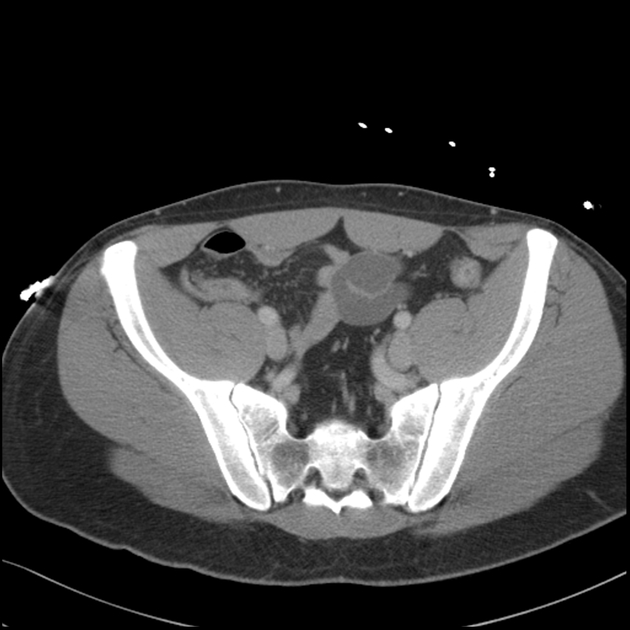
Muscle anatomy is again well seen, including iliopsoas muscle, gluteus maximus muscle, and obturator internus muscle (arrowhead). The muscles of the pelvis, hip and buttock anatomical chart. Functional anatomy of the male pelvicfloor. ► shoulder ► elbow ► wrist ► finger ► thumb. This section of the website will explain large and minute details of axial male pelvis use the mouse scroll wheel to move the images up and down alternatively use the tiny arrows (>>) on both side of the image to move the images. The lateral superficial muscles, the transversus and external and internal oblique muscles, originate on the rib cage and on the pelvis (iliac crest and inguinal ligament) and are attached to the anterior and posterior layers of the sheath of the rectus. The levator ani muscle, also known as the muscular pelvic diaphragm, is the musculotendinous sheet that forms the majority of the pelvic floor, supports the pelvic viscera, and aids in urinary and fecal evacuation as well as maintaining continence. The pelvic region holds major organs under its layers of muscles. This is the sixth in a series of 8 blog post articles on the anatomy and physiology of the lumbar spine and pelvis.
The pelvic region holds major organs under its layers of muscles.
Anatomic relationship between the vaginal apex and the bony architecture of the pelvis: Muscles of the pelvis that cross the lumbosacral joint to attach onto the trunk were described in the previous blog post note: Knowledge of normal pelvic anatomy on mri is critical for proper interpretation, in particular the standard visceral organ appearances, commonly encountered variants, and pathology mimics. We'll explore the structure of the parts, the difference between a male and female pelvis, and how to simplify the structure to make it manageable to draw. Functional anatomy of the male pelvicfloor. There are many muscles that form the pelvic floor, including puborectalis, pubococcygeus, iliococcygeus and coccygeus. Multisystem selenoprotein deficiency trunk common: Not only mri pelvis muscle anatomy, you could also find another pics such as pelvic mri anatomy, female pelvic mri anatomy, female pelvis mri axial, pelvic muscles. Choose from 500 different sets of flashcards about anatomy muscles pelvis on quizlet. Anatomical drawing of the female pelvis.
Posted by kenneth on november 18, 2019 anatomy muscles pelvis. The levator ani muscle, also known as the muscular pelvic diaphragm, is the musculotendinous sheet that forms the majority of the pelvic floor, supports the pelvic viscera, and aids in urinary and fecal evacuation as well as maintaining continence.

Posting Komentar untuk "Pelvis Muscles Mri Anatomy"